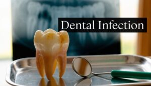
A Clearer, Safer Smile: Why We Chose Digital X-Rays for Our Vienna Patients
If you’ve ever had dental X-rays, you probably remember the old routine. You bit down on a rigid piece of film, tried not to move, and then waited while someone stepped away to process the image. For many patients, that process felt slower, less comfortable, and a little more stressful than it needed to be.
You may also have wondered about radiation exposure, especially if you bring in your children regularly, need follow-up imaging, or feel nervous about any dental procedure. Those concerns are reasonable. Good dental care should answer them directly, not brush them aside.
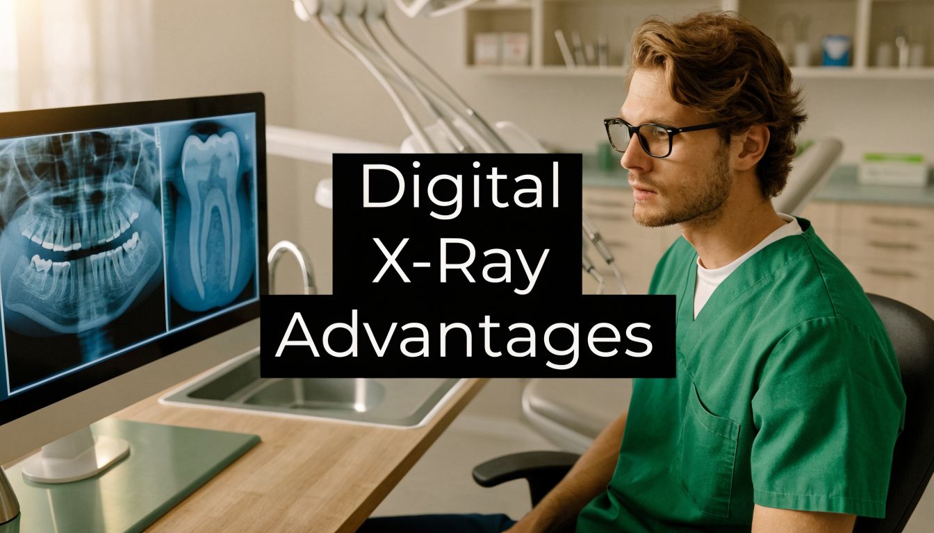
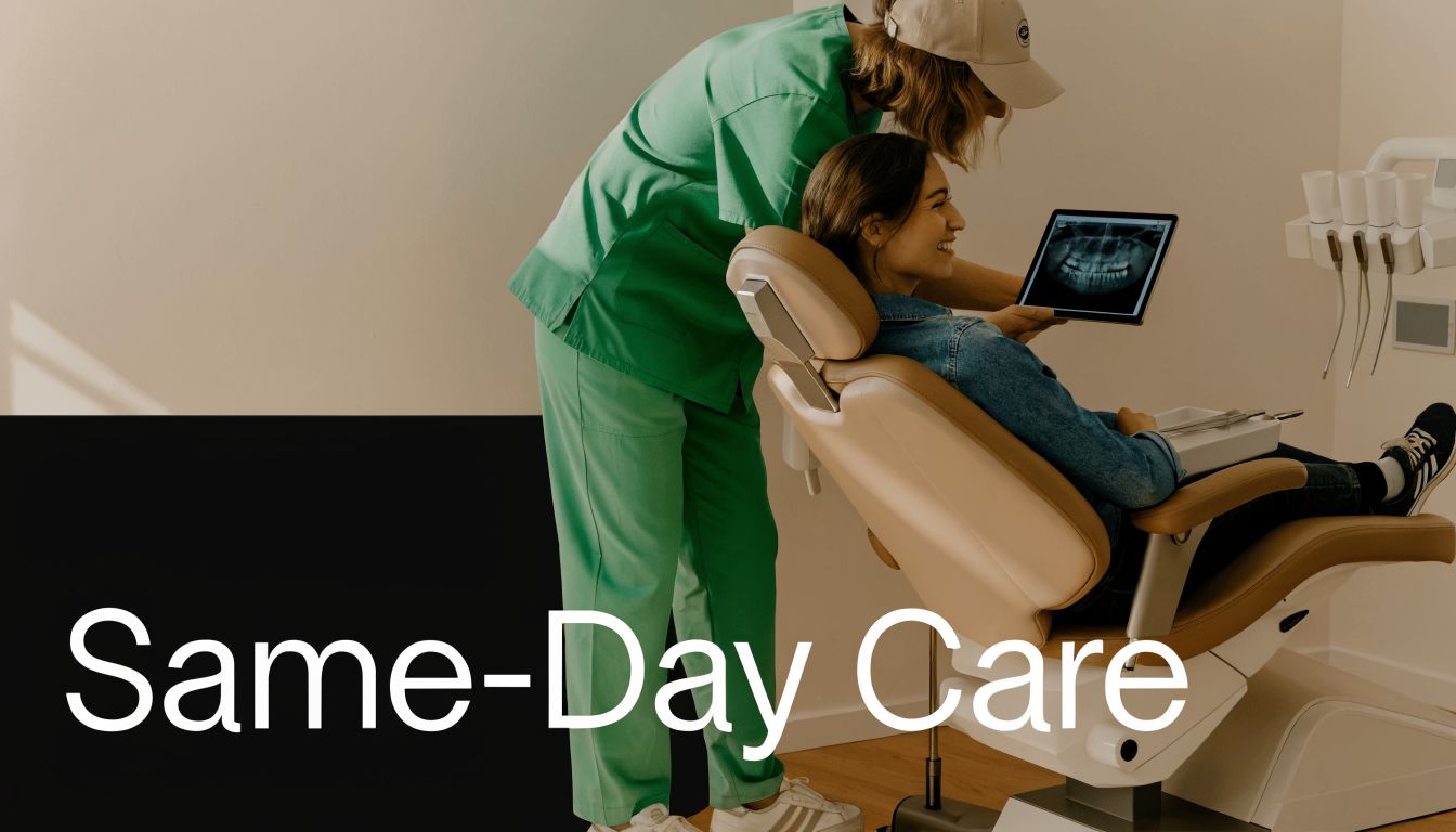
At Vienna Implant and Family Dentistry, we use digital X-rays because they make diagnosis faster, safer, and easier to understand. Dr. Vikram Chauhan chose this technology to support the kind of care patients in Vienna, VA, expect from a modern family and implant practice. That includes preventive checkups, same-day emergency visits, cosmetic planning, and complex restorative work.
Digital imaging also changes the feel of an appointment. Instead of waiting for film to be developed, patients can see clear images right away on a screen. That helps us explain what’s going on, whether we’re monitoring a child’s dental development, checking a painful tooth, planning a same-day crown, or evaluating bone support for implants.
Below are the digital x-ray advantages that matter most in real patient care. These aren’t abstract features. They affect how quickly we can find a problem, how comfortably you move through treatment, and how confidently we can plan care for your long-term oral health in Northern Virginia.
1. Reduced Radiation Exposure
For many patients, this is the first question. Is digital imaging safer than older film X-rays?
Yes. Digital X-rays reduce radiation exposure by 70% to 90% compared with traditional film-based X-rays, according to this overview of digital X-ray safety benefits in dentistry. That matters in any dental office, but it matters even more in family dentistry where children, pregnant patients, and people who need periodic follow-up imaging all deserve a cautious approach.

Lower exposure doesn’t mean lower usefulness. The sensors used in digital radiography are more responsive, so we can capture the detail we need with far less radiation than older film systems required. In practical terms, that helps us stay conservative while still getting the information needed to diagnose cavities, evaluate bone levels, or check healing after treatment.
Why this matters in everyday family care
This advantage is especially meaningful for routine checkups. A child who needs periodic monitoring, a patient in orthodontic or restorative care, or an adult preparing for implant treatment may need imaging more than once over time. Lower radiation helps reduce cumulative exposure over repeated scans.
It also helps anxious patients feel more at ease. When someone is already nervous about treatment, knowing the imaging process is designed to minimize exposure often makes the appointment feel more manageable.
Practical rule: If a technology lets us gather the same diagnostic information with less exposure, that’s the better choice for long-term care.
In our office, this supports a wide range of visits:
- Pediatric checkups: Children often need monitoring as teeth erupt and change.
- Pregnancy-related dental concerns: When imaging is clinically necessary, lower-dose digital systems support a safer approach.
- Implant and reconstruction planning: Some cases require more than one image over time, so dose reduction matters.
- Emergency care: We can evaluate the problem without adding unnecessary delay or repeat exposure.
What doesn’t work is assuming every image should be taken casually just because the dose is lower. Good dentistry still follows judgment, timing, and need. Digital technology improves safety, but it doesn’t replace clinical decision-making. It strengthens it.
2. Improved Image Quality and Diagnostic Clarity
A sharper image changes what we can catch early. That’s one of the most useful digital x-ray advantages in everyday practice.
Digital X-rays appear immediately and can be adjusted on screen for contrast, brightness, and magnification, which helps dentists detect subtle issues such as small cavities, infections, or abnormalities more clearly, as described in this review of digital X-ray image quality and immediate display. Instead of relying on a fixed film image, we can enlarge an area, improve contrast, and study details chairside with the patient.
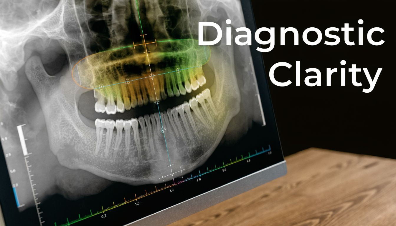
That clarity matters in cases that don’t look dramatic from the outside. A patient may only feel mild sensitivity, but a clearer image can reveal a cavity between teeth, hidden infection, or bone changes that explain the symptoms. Better imaging helps us make decisions earlier, when treatment is often simpler.
Where image clarity changes treatment
This is especially important in restorative and implant dentistry. When planning treatment, small details affect the final result. We need to see tooth structure, root shape, existing restorations, and supporting bone as clearly as possible.
For patients who need more advanced evaluation, digital imaging also works well alongside 3D dental imaging technology. That combination helps when we’re assessing implant sites, bone contours, or complex restorative needs in greater detail.
A few common examples:
- Early decay detection: A tiny area between teeth may not be visible in a mirror exam.
- Bone level evaluation: Gum disease and tooth support are easier to track when image detail is consistent.
- Cracked or failing restorations: A better view helps distinguish a minor issue from a tooth that needs more extensive treatment.
- Implant planning: Clear visualization supports more precise decisions before treatment starts.
Clear images don’t just help the dentist. They help the patient understand why a recommendation makes sense.
What doesn’t work is using good images without good interpretation. Technology is a tool, not a diagnosis by itself. The primary benefit comes from pairing image quality with a careful exam, symptoms, and experience. When those pieces line up, treatment recommendations become more accurate and easier for patients to trust.
3. Faster Workflow and Same-Day Treatment Capability
Speed matters when you’re in pain. It also matters when you’re trying to fit dentistry into a workday, school schedule, or busy family calendar.
Digital radiography supports that pace because the image shows up almost immediately. In the broader digital X-ray market, direct radiography systems held approximately 65% share in 2023, reflecting strong adoption of systems that deliver immediate images and streamline workflow in settings such as hospitals and dental clinics, according to this report on the global digital X-ray systems market.
For patients, the practical benefit is simple. We don’t have to stop the visit and wait on film processing. We can take the image, review it right away, and decide on the next step while you’re still in the chair.
Why speed matters for real appointments
This is one of the reasons digital imaging fits so well with emergency care and restorative treatment. If a patient comes in with a broken tooth, swelling, or sharp pain, immediate imaging helps us identify the cause and move quickly toward relief. The same applies when someone wants efficient restorative care instead of multiple drawn-out visits.
That workflow supports services like same-day CEREC crowns in a single visit. A fast, accurate image is part of what allows diagnosis, planning, and treatment to happen more smoothly in one appointment.
What works and what doesn’t
What works is using that speed to improve care, not to rush it. Fast imaging gives us more time for explanation, treatment planning, and actual dentistry. It shouldn’t mean cutting corners.
A few scenarios where patients notice the difference most:
- Emergency visits: Faster diagnosis means quicker pain relief planning.
- Same-day crown appointments: Immediate images support a more efficient restorative workflow.
- Routine family visits: Less waiting often means a smoother appointment for children and parents.
- Treatment changes mid-visit: If something unexpected appears, we can respond right away.
What doesn’t work is assuming every fast process is automatically a better process. If a sensor is positioned poorly or the patient moves, the image still needs to be usable. Training and attention to detail matter. Digital systems save time best when the clinical team uses them well.
4. Enhanced Patient Communication and Education
A lot of dental anxiety comes from not knowing what the dentist sees. Patients hear words like cavity, bone loss, infection, or crown, but without a visual reference, those terms can feel abstract.
Digital X-rays change that conversation. When the image appears on a screen beside the chair, we can point directly to the tooth, root, filling, or area of concern. That makes the diagnosis easier to follow and gives patients a clearer reason for treatment recommendations.
Seeing the problem often lowers anxiety
This is particularly helpful with children, teens, and adults who’ve put off care because they’re worried about what they might hear. When a parent can see where decay is starting, or a patient can see the outline of a cracked tooth, the discussion becomes less mysterious. It turns into a practical conversation about next steps.
We use that visual approach in many kinds of appointments:
- Preventive visits: Showing early wear, developing decay, or changes over time.
- Periodontal discussions: Helping patients understand why gum support matters.
- Restorative planning: Explaining why a filling may be enough in one case, but a crown is better in another.
- Implant consultations: Showing available bone and nearby structures so treatment feels more concrete.
“When patients can see the issue, they usually ask better questions and feel more confident about the plan.”
Digital images also help us compare old and new findings. That’s useful when monitoring a tooth over time or explaining why treatment that could once wait should now move forward. Patients often appreciate that we’re not relying on vague language. We can show them the progression.
Better communication leads to better follow-through
In real practice, understanding improves compliance. A patient who sees a small cavity developing is more likely to accept a conservative filling before that tooth turns into an emergency. A parent who sees crowding or eruption concerns is more likely to keep regular recall visits. A patient considering cosmetic or restorative treatment can better understand how function and appearance connect.
What doesn’t work is overwhelming patients with technical language. Digital tools are excellent, but explanation still needs to stay clear and calm. A good chairside conversation should leave the patient informed, not confused. That’s where technology and communication need to work together.
5. Seamless Integration with Digital Workflows and Practice Management
Some digital x-ray advantages aren’t obvious to patients at first, but they still shape the quality of the visit. One of the biggest is how well digital imaging fits into a fully modern dental workflow.
Instead of creating a physical film that has to be handled, stored, or manually moved from one step to the next, digital images can be stored electronically and shared as part of a connected patient record. That matters because dentistry rarely happens in isolation. Imaging, exams, treatment planning, restorative design, and follow-up all need to line up.
Why integration matters behind the scenes
When an image is easy to retrieve, compare, and review, appointments become more organized. If a patient returns for a crown check, emergency visit, implant consultation, or cosmetic discussion, the diagnostic record is already part of the broader picture. That makes continuity of care easier.
In a practice offering preventive, restorative, implant, and cosmetic services under one roof, that connected workflow supports smoother care in several ways:
- Treatment planning stays organized: Images remain linked to the patient’s ongoing record.
- Referrals and collaboration are easier: Digital files are simpler to review and share when needed.
- Progress tracking is clearer: Comparing images over time supports long-term monitoring.
- Patient conversations are more efficient: We can pull up prior findings instead of starting from scratch.
This matters in emergency dentistry, too. If a patient has seen us before and returns with a new issue, prior digital records help us assess what changed. That can make decision-making quicker and more precise.
Practical value in a modern dental office
Patients don’t usually choose a dentist because of software integration, but they do notice the results. Fewer delays. Less repeated explanation. More consistent records. A smoother handoff from diagnosis to treatment.
What works is using digital systems to reduce friction. What doesn’t work is adding technology that creates more clicking, more confusion, or less patient interaction. The point isn’t to make the office feel mechanical. The point is to free up time and attention for actual care.
In practice: The best digital workflow is the one patients barely notice, because everything simply feels more coordinated.
That’s a key advantage. When imaging fits naturally into the rest of the appointment, patients experience a visit that feels calmer, clearer, and more efficient.
6. Cost Efficiency and Long-Term Savings
Patients don’t always ask about the business side of digital imaging, but it still affects the care experience. A system that removes waste, cuts delays, and simplifies storage helps a practice invest time and resources more wisely.
Digital X-rays eliminate film, chemical processing, and physical image storage. That doesn’t mean the equipment is inexpensive at the start. It usually isn’t. The upfront investment is one of the key trade-offs, especially for smaller practices deciding when to upgrade.
The trade-off is upfront cost versus ongoing efficiency
Traditional film systems create recurring costs. You need film, processing supplies, storage space, and the staff time that goes with handling all of it. Digital imaging removes many of those recurring burdens, which is one reason it has become such a standard part of modern care.
For patients, the benefits show up indirectly but clearly:
- Appointments move more efficiently: Less waiting and less back-and-forth.
- Records are easier to retrieve: Old images don’t disappear into physical archives.
- Clinical time is used better: The team spends more time on diagnosis and treatment, not processing film.
- Practice resources can support other technology: Savings in one area can help a practice continue modernizing.
There’s also less waste built into the process. No darkroom setup. No film developing chemicals. No stacks of physical radiographs to manage over time.
What works for patients
From a patient perspective, the best use of digital technology is practical. It should improve convenience and support better care. If a practice buys digital equipment but doesn’t train staff well or doesn’t build efficient systems around it, patients won’t feel much benefit.
That’s why implementation matters as much as the equipment itself. A good digital workflow reduces bottlenecks. It helps the team move confidently from image capture to diagnosis, explanation, and treatment. In a busy office serving families, emergency patients, and restorative cases, that consistency matters every day.
What doesn’t work is treating “digital” as a marketing label by itself. The true value lies in how well the office uses the technology. When used properly, digital X-rays support efficiency that patients can experience in shorter waits, clearer communication, and more organized care.
7. Advanced Implant Planning and Surgical Precision
Implant dentistry is one area where imaging quality directly affects treatment planning. If you’re replacing a missing tooth, or several teeth, the question isn’t just where a tooth should go. It’s whether the underlying bone can support it, how nearby structures affect placement, and how to build a plan that’s predictable.
That’s why digital imaging is so important in implant cases. It gives us a clearer foundation for evaluating the site before any procedure begins. For simple cases, standard digital X-rays may provide key information. For more complex planning, digital radiography often works alongside 3D imaging to assess anatomy in greater detail.
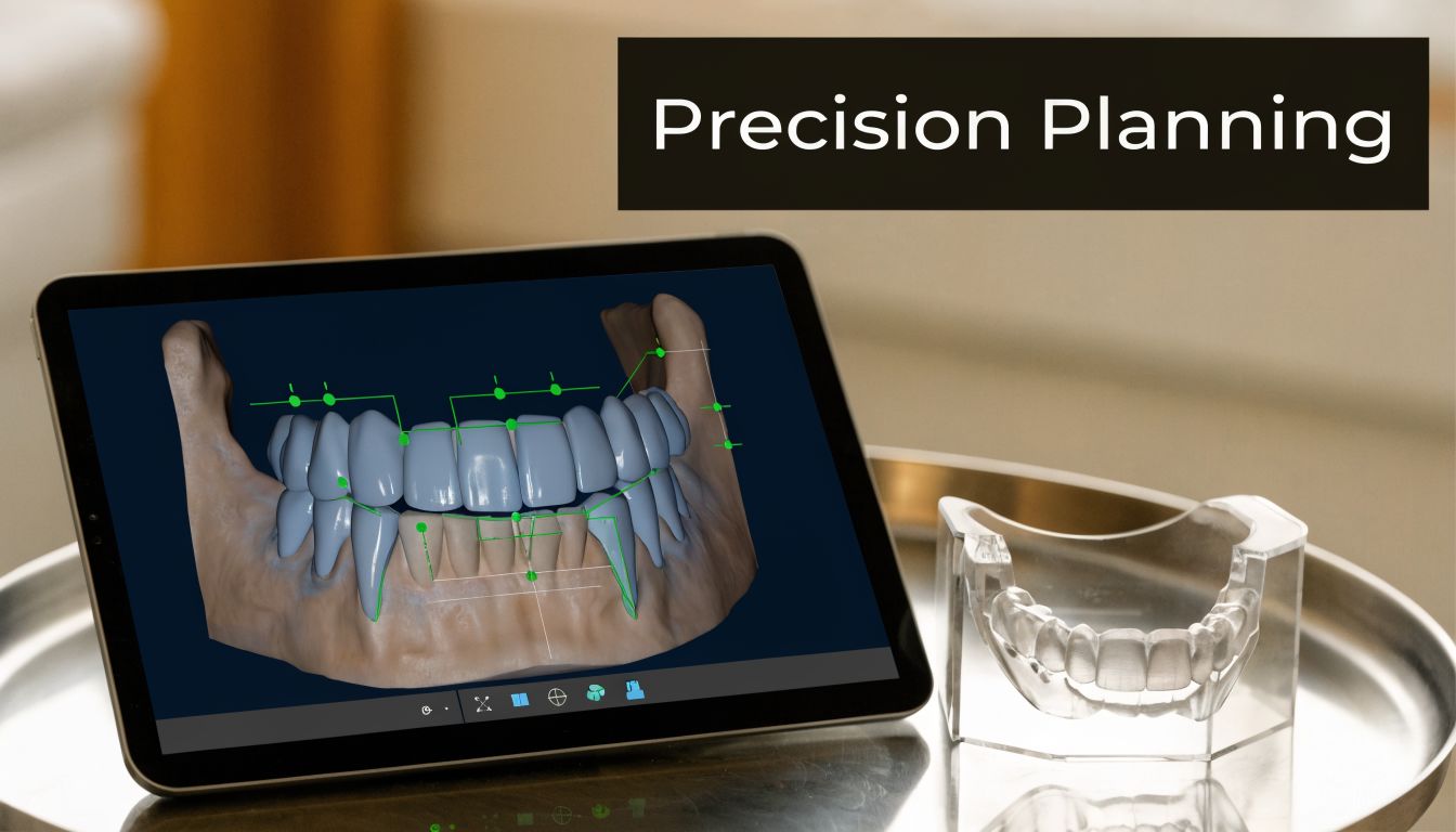
Precision matters before treatment starts
A patient may look at a missing tooth and think the solution is straightforward. Sometimes it is. Sometimes the supporting bone has changed, the space is limited, or the site needs additional preparation first. Good imaging helps us identify those details early instead of discovering them halfway through treatment.
That’s especially relevant if a patient may need procedures such as bone grafting before dental implants. Digital imaging helps evaluate support, spacing, and surrounding structures so the plan matches the biology, not just the appearance of the gap.
Why this improves outcomes and confidence
Patients often feel more confident when they can see that implant planning is precise rather than approximate. That matters whether someone needs a single implant, multiple implants, or a broader full-mouth reconstruction plan. Imaging supports decisions about placement, restoration design, and timing.
This level of planning is also important in cosmetic and functional cases. If an implant supports a highly visible part of the smile, small positioning decisions can affect both appearance and bite. Better imaging helps reduce guesswork.
A few situations where this becomes especially valuable:
- Single-tooth implant replacement: Accurate site evaluation supports a more natural final result.
- Multiple missing teeth: Planning has to account for spacing, support, and restoration design.
- Teeth-in-a-Day and larger reconstructions: Complex cases benefit from detailed imaging before treatment begins.
- Sites with past bone loss or old dental work: Precision matters more when conditions are less straightforward.
Good implant treatment starts long before placement day. It starts with seeing the site accurately and planning conservatively.
What doesn’t work is promising precision without proper imaging. Implant dentistry depends on planning. Digital X-rays help create that plan in a way that is safer, clearer, and easier for patients to understand.
8. Environmentally Responsible and Sustainable Dentistry
Most patients think about dental X-rays in terms of safety and speed. Fewer people think about waste, but it’s still worth discussing.
Traditional film X-rays depend on physical materials and chemical processing. Digital systems remove that part of the workflow. They eliminate the need for film development chemicals and reduce the storage burden that comes with paper-heavy, film-heavy systems. That makes digital imaging a cleaner choice for a modern dental office.
A practical environmental advantage
This isn’t the kind of benefit that changes a diagnosis on its own, but it does reflect how a practice operates. Patients increasingly want healthcare that feels current, efficient, and responsible. Digital imaging supports that by reducing waste associated with old film-based methods.
There’s also a practical office benefit. When a team doesn’t need to process film or manage chemical disposal, the workflow becomes simpler and the environment becomes easier to maintain. That supports a cleaner, more efficient clinical setting.
These environmental advantages are part of why digital imaging has become standard in so many care environments. In the broader market, hospitals have been moving rapidly from analog to digital systems, with that transition projected at 4.5% CAGR through 2031, according to this analysis of the digital X-ray market and analog-to-digital transition. Dentistry benefits from the same general shift toward cleaner, faster, digitally managed imaging.
What this means for patients in Vienna
For local families, this benefit is less about marketing and more about alignment. A modern dental office should be thinking about efficiency, safety, and waste reduction together. Digital X-rays support all three.
- No film chemicals: That removes an older source of clinical waste.
- Less physical storage: Images can be retained electronically instead of in bulky archives.
- A more efficient office: Fewer manual steps often mean fewer delays and less clutter.
- A better fit with digital care overall: Imaging, records, and treatment planning all work together.
What doesn’t work is treating sustainability as a substitute for clinical quality. Patients still need accuracy first. The environmental benefit matters because it comes alongside better workflow and better imaging, not instead of them.
Digital X-Ray: 8 Advantages Compared
| Item | Implementation Complexity 🔄 | Resource Requirements ⚡ | Expected Outcomes ⭐ | Ideal Use Cases 📊 | Key Advantages & Tips 💡 |
|---|---|---|---|---|---|
| Reduced Radiation Exposure | Low–Moderate: simple sensor setup; staff positioning training | Moderate: digital sensors, workstation, initial capital | ⭐ High: markedly lower patient dose; safer for repeat imaging | Pediatric care, pregnant patients, routine family monitoring | Educate patients on dose reduction; train staff; document doses |
| Improved Image Quality and Diagnostic Clarity | Moderate: requires calibrated displays and operator skill | Moderate–High: high-res sensors, quality monitors | ⭐⭐ Very High: earlier detection and precise diagnoses | Implant planning, full‑mouth reconstruction, early caries detection | Calibrate monitors regularly; use enhancement tools; archive images |
| Faster Workflow and Same-Day Treatment Capability | Moderate: software integration and workflow redesign | Moderate: networked systems, tablets, CAD/CAM links | ⭐ High: reduced chair time; enables same‑day treatments | CEREC crowns, emergency visits, same‑day implant decisions | Integrate with practice software; train staff; maintain backups |
| Enhanced Patient Communication and Education | Low: display setup and clinician communication skills | Low–Moderate: chairside monitors/tablets, annotation tools | ⭐ High: improved acceptance, reduced anxiety, better compliance | Pediatric consults, implant consent, patient education sessions | Use simple language; position screens for visibility; save annotated images |
| Seamless Integration with Digital Workflows and Practice Management | High: EHR/CAD/CAM and imaging interoperability | High: IT infrastructure, cloud storage, licenses, training | ⭐⭐ High: streamlined admin, accurate records, faster planning | Comprehensive implant workflows, teleconsults, CEREC integration | Choose compatible systems; enforce backups/security; train staff |
| Cost Efficiency and Long-Term Savings | Moderate: planning ROI and phased adoption | High upfront; low ongoing consumables and maintenance | ⭐ High (long term): reduced consumable costs; improved profitability | Growing practices investing in tech and implant services | Calculate TCO; consider lease vs purchase; track ROI metrics |
| Advanced Implant Planning and Surgical Precision | Very High: CBCT use, 3D planning software, specialist training | Very High: CBCT units, planning software, guide fabrication | ⭐⭐⭐ Exceptional: precise placement, fewer complications | Complex implant cases, Teeth‑in‑a‑Day, full‑mouth reconstruction | Invest in surgical guides; document plans; collaborate with specialists |
| Environmentally Responsible and Sustainable Dentistry | Low: eliminates darkroom processes | Low–Moderate: digital equipment plus e‑waste management | ⭐ Moderate: reduced chemical waste and paper use; lower compliance burden | Practices marketing sustainability; community health initiatives | Track waste reduction metrics; implement e‑waste recycling; promote benefits |
Experience the Difference at Vienna Implant and Family Dentistry
At Vienna Implant and Family Dentistry, technology only matters if it makes care better for the patient sitting in the chair. That’s the standard we use when we choose equipment and build our workflow. Digital X-rays meet that standard because they support safer imaging, faster answers, and clearer communication during every kind of visit.
For routine family dentistry, that means a more comfortable and efficient experience. If your child needs a checkup, or if you’re staying consistent with preventive care as an adult, digital imaging helps us gather the information we need without the delays and inconvenience that older film systems created. Patients spend less time waiting and more time talking through what their teeth need.
For emergency appointments, the difference is even more obvious. When someone arrives with a broken tooth, swelling, or pain that can’t wait, immediate digital images help us identify the problem quickly and start discussing relief right away. That’s one reason modern imaging matters so much in a practice that sees urgent dental concerns and same-day needs.
For restorative and cosmetic care, digital X-rays help us plan with more confidence. If you need a filling, crown, bridge, or more extensive treatment, detailed images support better decision-making from the start. If you’re considering same-day CEREC treatment, the ability to review images immediately helps keep the appointment focused and efficient. If you’re exploring veneers or a smile makeover, imaging also helps confirm that the foundation of the smile is healthy enough to support cosmetic work.
Implant patients often feel the biggest impact. Replacing missing teeth requires more than just choosing the right restoration. It requires understanding the bone, the spacing, the bite, and the long-term plan. Digital imaging helps us evaluate those details carefully, and when more advanced imaging is needed, it becomes part of a more complete planning process. That can make treatment feel more understandable and less intimidating.
Just as important, digital X-rays help patients feel involved. When Dr. Vikram Chauhan can bring your image up on a screen and point directly to the area in question, the conversation changes. You’re not being asked to trust a vague explanation. You can see the issue for yourself, ask questions, and understand why a certain treatment makes sense. For anxious patients, that kind of transparency often reduces a lot of uncertainty.
There are trade-offs with any technology. Digital systems require investment, training, and a team that knows how to use them well. The benefit comes from using the technology thoughtfully, not just having it in the office. That’s why the patient experience matters so much. The best digital tools should make your visit feel smoother, not more complicated.
If you’re looking for a dentist in Vienna, VA, who uses modern imaging as part of practical, patient-centered care, Vienna Implant and Family Dentistry is one local option to consider. Our office is located at 112 Pleasant St. NW, Suite H, Vienna, VA 22180. If you’re ready to schedule a consultation for preventive care, dental implants, same-day crowns, emergency treatment, or cosmetic dentistry, contact Vienna Implant and Family Dentistry and experience a more modern approach to dental care.